
A New Study in 495 Centenarians Points to a Brain Aging Biomarker You Probably Have Not Heard Of
Most people know that high blood pressure predicts heart disease and high blood sugar predicts diabetes. The equivalent toolkit for brain aging has been slower to arrive, but it is now here. Modern assessments combine brain MRI volumetrics, cognitive testing, genetic analysis, and blood biomarkers to estimate how fast your brain is aging and what is driving the trajectory. Within the blood biomarker panel, one marker has been quietly building evidence for a decade as a particularly powerful indicator. A study published this week in JAMA Network Open just gave it a major boost.
The study followed 495 Japanese centenarians, people aged 100 and older, for up to 17 years. Researchers measured three blood markers at the start, the two most associated with Alzheimer’s disease (amyloid-beta and phosphorylated tau) and a third called neurofilament light chain, abbreviated NfL. NfL outperformed both Alzheimer’s-specific markers as a predictor of both cognitive function and lifespan, with each one standard deviation increase corresponding to a 36% higher mortality risk after adjustment for age, sex, kidney function, and other health factors.
NfL has been gaining ground as one of the more informative blood biomarkers in brain aging research, and this study adds to the case for paying attention to it.
What NfL is in plain language
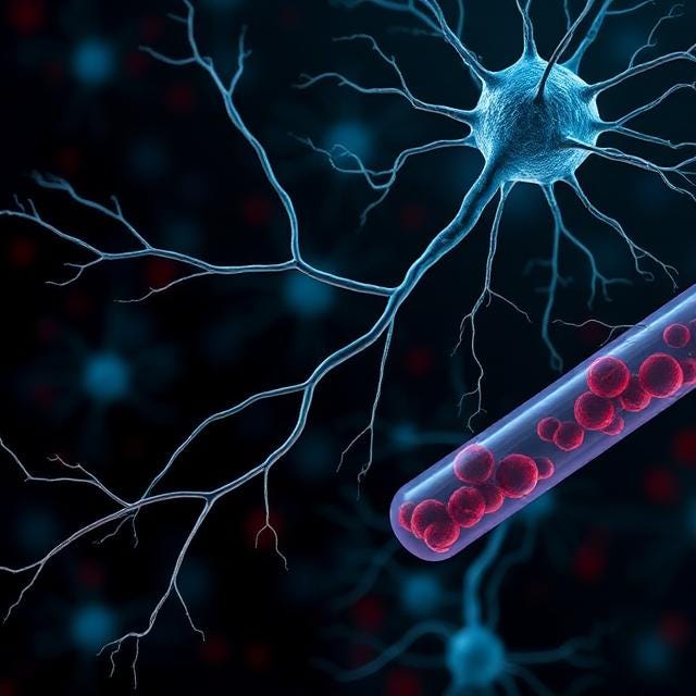
Your nervous system is a network of long, thin cells called neurons. The signal-carrying part of each neuron is called an axon, and axons can be very long. A single axon running from your spinal cord to your foot is about a meter in length. To stay structurally intact across that distance, axons rely on an internal scaffolding made of proteins. One of those proteins is neurofilament light chain.
When axons get damaged from any cause, whether from an injury, an autoimmune attack, a stroke, or the slow wear and tear of aging, fragments of NfL leak out of the cell and into the bloodstream. Once in the blood, NfL can be measured with ultrasensitive assays. The level reflects how much axonal damage is happening across the entire nervous system.
A useful way to think about NfL is as a smoke detector for nerve damage. It does not tell you exactly where the damage is or what is causing it. It does tell you whether the damage is happening, and how fast.
The level measured in any given NfL blood draw reflects two things layered together. There is a chronic baseline that drifts upward with age and accumulated nervous system wear, and there is an acute component that rises and falls in response to recent injuries or active disease. A reading taken a month after a concussion will look different from one taken a year later, even if nothing else has changed. This is why interpreting NfL is most useful with both age-stratified reference ranges, which anchor the chronic component, and serial measurements over time, which separate the underlying trajectory from short-term spikes.
Why NfL is gaining attention
For most of the last 20 years, the dominant story in dementia research has been about amyloid plaques and tau tangles, the two protein deposits that define Alzheimer’s disease pathology. Drugs targeting amyloid have now been approved (lecanemab and donanemab), and they do clear plaques. The cognitive benefits have been modest, and the conversation in the field has shifted toward whether amyloid and tau are the main drivers of brain aging in most people, or whether something broader is at work.
NfL captures that broader picture. It rises when axons are damaged for any reason. The contributors include the small vessel disease that quietly accumulates in everyone’s brain over decades, the chronic inflammation that follows infections and metabolic stress, and the cellular wear of normal aging. For people who reach extreme old age, where multiple subtle pathologies coexist, the centenarian study suggests NfL is what tracks brain trajectory and lifespan most closely.
A second study, from Dr. Mathias Jucker’s group at the German Center for Neurodegenerative Diseases, published in PLOS Biology in February 2026, extends the picture across species. NfL rises with age in mice, dogs, cats, and horses, and it predicts mortality in those species too. NfL is one of the very few blood biomarkers that works the same way across mammals, and that kind of evolutionary conservation is a strong sign that NfL is reflecting something fundamental about biological aging rather than a quirk of human disease.
Why someone might want to know their NfL level
The case for measuring NfL comes down to four practical points.
First, NfL responds to injuries and interventions on a timescale of weeks. After a traumatic brain injury or concussion, plasma NfL rises within days, peaks at roughly 10 days to 6 weeks, and can remain elevated for months (Shahim et al., Sci Transl Med 2021). This response curve is part of why serial NfL measurements are now being incorporated into recovery tracking after head injuries, including in sports medicine where biomarker trajectories are starting to inform return-to-play decisions in concussed athletes (McDonald et al., JAMA Netw Open 2024). After a multiple sclerosis relapse, NfL peaks at 3 to 4 weeks, and treatment effects on NfL typically show up within 3 to 6 months in MS clinical trials. MRI volume changes from interventions can also be detected within 3 to 4 months in some studies, particularly in clinical populations like mild cognitive impairment, but NfL gives a faster individual-level readout on whether the underlying damage process is responding to whatever someone is doing.
Second, NfL is more accessible than MRI for repeat monitoring. A direct-to-consumer NfL test runs roughly $200 to $400, compared with $600 to $8,000 for a brain MRI. A blood draw also works for people who cannot easily get into an MRI scanner, such as those with pacemakers or significant claustrophobia. For someone considering serial monitoring once or twice a year, the difference in cost and convenience adds up.
Third, NfL is non-specific in a useful way. The criticism sometimes leveled at NfL is that it does not diagnose any single disease, and that is correct. It is not an Alzheimer’s test or an MS test. What it does measure is the overall rate of nerve damage in your body. For a person who wants a single number to track brain health over time, that breadth is a feature rather than a flaw.
Fourth, the value of NfL is in the trajectory, not the snapshot. A single elevated NfL does not mean disease, and a single normal NfL does not mean safety. What matters is how your level compares to age-matched ranges and how it changes over time. People who track their NfL annually can see whether their brain aging is accelerating, holding steady, or slowing in response to whatever they are doing.
What raises NfL
Several things drive NfL up, and most of them will be familiar from cardiovascular risk lists. Age itself is the largest single factor, with levels roughly doubling between age 30 and age 70 in healthy populations, which is why reference ranges have to be age-adjusted to be interpretable. Acute neurological events including stroke, traumatic brain injury, multiple sclerosis flares, and concussion produce sharp spikes that can take weeks to months to come back down.
Chronic conditions push NfL up gradually rather than acutely. Uncontrolled hypertension, diabetes, sleep apnea, chronic kidney disease, and longstanding low-grade inflammation are the usual contributors, since each one quietly damages the brain over time. Two non-disease factors are also worth knowing about. Vigorous exercise produces a transient rise in NfL lasting up to 72 hours, so the timing of a blood draw matters and a recent hard workout will distort the reading. Body size affects NfL through dilution, since larger blood volume produces lower concentrations of any given protein, and obesity has been associated with lower NfL values even when underlying brain damage is comparable.
What lowers NfL based on existing evidence
The cleanest signal comes from drugs that turn off an active driver of nerve damage.
In a rare form of ALS caused by mutations in the SOD1 gene, the antisense drug tofersen reduced plasma NfL by approximately 60% over 28 weeks compared with placebo (Miller et al., NEJM 2022). This was the first time the FDA accepted a blood biomarker like this as a surrogate endpoint for a neurological drug.
In multiple sclerosis, several modern disease-modifying therapies, including ocrelizumab, ofatumumab, siponimod, and fingolimod, consistently lower serum NfL alongside their effects on relapse rates and brain lesions.
For people without an active neurological disease, the data on lifestyle interventions is more mixed but encouraging in places. A pilot randomized trial of 36 women with MS found that a Mediterranean dietary pattern significantly reduced plasma NfL over six months versus their usual diet (Verter et al., Neurology 2024). A 2025 cohort analysis of approximately 1,500 older adults including both healthy individuals and people with Alzheimer’s disease found lower plasma NfL levels in those with higher MIND or Mediterranean diet adherence (Liu et al., Sci Rep 2025).
Exercise data is mixed in healthy populations and people with Alzheimer’s, but more consistently positive in MS, where structured moderate-intensity aerobic and resistance work has reduced NfL in several trials.
Two recent results push the other way. In Phase 3 trials of oral semaglutide for early Alzheimer’s, NfL went up slightly even as some other markers went down, and the trial missed its cognitive endpoints. The amyloid-clearing drug lecanemab, which has FDA approval for early Alzheimer’s, reduced amyloid and tau markers but did not significantly reduce NfL in its pivotal trial.
NfL responds to interventions that address the underlying axonal damage, not necessarily to interventions that lower a related but distinct marker. Alzheimer’s and related neurodegenerative conditions have multiple contributing factors, and amyloid plaques and tau tangles are only part of the story. NfL captures damage from aging itself, from cerebrovascular contributors, and from injury, all of which influence cognitive trajectories alongside the classical AD pathologies. Combined with MRI volumetrics, plasma pTau217, and the amyloid-beta 42/40 ratio, NfL contributes to a more complete picture of what is driving an individual’s brain aging. NfL is being incorporated as a complementary endpoint in modern trial design rather than a replacement for the markers that came before it.
Where the test is available
Plasma NfL is now offered by several major commercial labs in the United States, although insurance coverage varies and most people will pay out of pocket.
Labcorp offers a serum NfL test (test number 140455), with age-stratified reference ranges. Quest Diagnostics offers a similar plasma NfL assay. Both are available with a physician order.
For people without easy access to a physician willing to order it, several direct-to-consumer testing services now offer NfL through CLIA-certified labs without a referral. Pricing for direct-to-consumer NfL tests typically runs between $200 and $400. The underlying assay is usually the same Quanterix Simoa technology used in research studies.
The Roche/Labcorp and Siemens automated platforms are now in clinical use in some countries. The Siemens assay was the first blood-based NfL assay to receive regulatory clearance in Europe for monitoring MS disease activity.
Two practical points are worth knowing for anyone considering testing. Results are most useful when interpreted against age-stratified reference ranges, since absolute values vary substantially across labs and platforms. Serial measurements over time in the same person at the same lab are also more informative than a single value in isolation.
At NeuroAge, NfL can be measured at the RNA level in our blood test alongside other genes selected for brain aging signal. We are not specifically giving NfL results at this time but are looking into doing this in the future. Our blood test is combined with brain MRI volumetrics, genetic resilience analysis, and cognitive testing in a multi-modal brain aging assessment.
Where this is heading
The next generation of clinical trials in brain aging will use plasma biomarkers like NfL to identify people for intervention before symptoms appear. This is already happening in rare genetic forms of ALS through the ATLAS trial, which uses plasma NfL as a trigger to start tofersen treatment in presymptomatic SOD1 mutation carriers. The same logic, biomarker-triggered preventive therapy, is being adapted for Alzheimer’s prevention through trials like DIAN-TU Primary Prevention and APEX. These are restricted to specific high-risk groups for now, but they are the template for what brain aging prevention will look like for everyone in the years ahead.
A second future direction worth flagging is the move toward predicting brain imaging findings directly from blood biomarker panels. Brain MRI captures information that no blood test currently replicates, including the regional pattern of atrophy and the burden of small vessel disease in white matter. MRI is also expensive and access remains uneven, which constrains how broadly imaging can be used for prevention. Several research groups are now training models that combine plasma markers (NfL, phosphorylated tau 217, GFAP, amyloid-beta 42/40) along with genetic and demographic factors to predict gray matter atrophy and white matter hyperintensity burden. In the Baltimore Longitudinal Study of Aging (622 cognitively unimpaired older adults followed for 4.7 years), baseline plasma pTau-181 and GFAP independently predicted longitudinal gray matter loss and ventricular enlargement, with NfL adding complementary information about ongoing axonal injury (Dark et al., Alzheimer’s Res Ther 2024). The eventual goal is a blood-based screening pathway that flags people whose trajectory warrants confirmatory imaging, rather than imaging everyone upfront. The platforms most likely to develop those predictive models will be the ones that already collect blood biomarkers, MRI, cognitive testing, and genetics on the same people over time, which is the data structure NeuroAge was built around.
The Shikimoto et al. centenarian paper makes a quieter but more universal point. Among people who have already won the longevity lottery by reaching 100, the marker that best predicts who will keep their cognition and who will not is the one that captures the broadest range of damage to the nervous system. That is exactly the kind of biomarker that belongs at the center of brain aging research and clinical practice. Knowing your level, and tracking it over time, is becoming a reasonable thing to consider for anyone who cares about how their brain ages.

Written by
Dr. Christin Glorioso, MD PhD
Dr. Glorioso is the founder and CEO of NeuroAge Therapeutics. With her background in neuroscience and medicine, she is dedicated to revolutionizing brain health and helping people maintain cognitive vitality.
Learn more about Dr. Glorioso

