

Fewer Than One in Five People With This Treatable Cause of Dementia Ever Get Diagnosed
 Dr. Christin Glorioso, MD PhD19 min read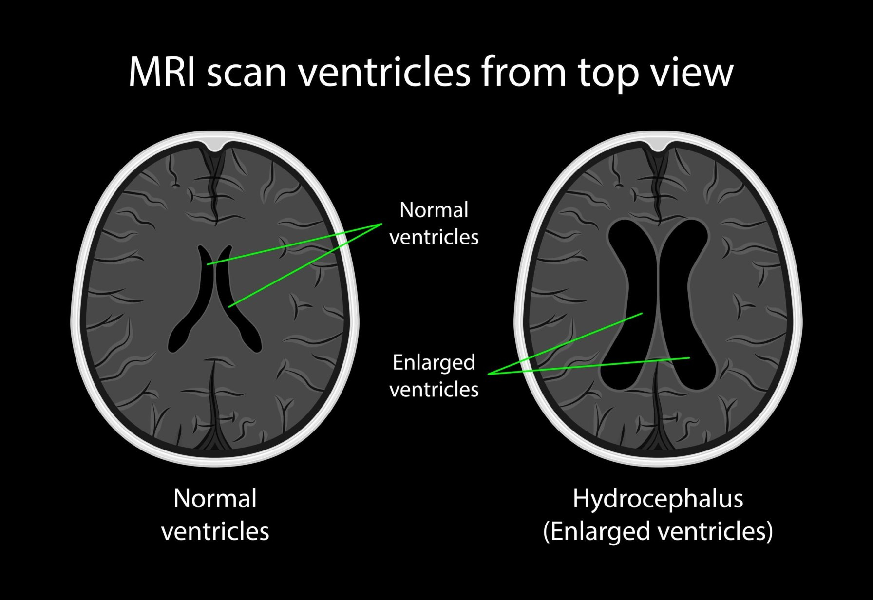
The Hydrocephalus Association estimates that close to 700,000 American adults have normal pressure hydrocephalus, and that fewer than 20 percent of them ever receive the diagnosis. The figure gets repeated by the Alzheimer’s Association, by academic medical centers, and in nearly every article written about the condition. A second claim usually accompanies it, which is that normal pressure hydrocephalus accounts for somewhere between one and six percent of all dementia cases and is one of the few forms of dementia that can be directly treated.
At NeuroAge, brain MRI is one of the four components of the brain aging assessment I built the company around. Enlarged ventricles show up on those scans with some regularity, in people who have no symptoms at all and in people who do have symptoms but haven’t connected them to their cause. Sometimes the ventricles are at the far end of the normal range for age. Sometimes there is white matter signal change alongside them. An open question for clients and the physicians treating them is what, if anything, should be done about enlarged ventricles?
That question turns out to sit on top of an unusual amount of unresolved science. This article covers what enlarged ventricles are, what they predict, why the diagnosis of normal pressure hydrocephalus depends on a convention rather than a biological test, whether treating the condition prevents dementia, and how the answer might change for someone planning to live past 100.
What the ventricles are and why they get bigger
The ventricles are four connected chambers inside the brain filled with cerebrospinal fluid, a clear liquid that cushions the brain, delivers nutrients, and carries waste away. The fluid is produced inside the ventricles, circulates around the brain and spinal cord, and is reabsorbed back into the bloodstream. During sleep this circulation participates in the brain’s overnight waste clearance system, which removes proteins including amyloid and tau.
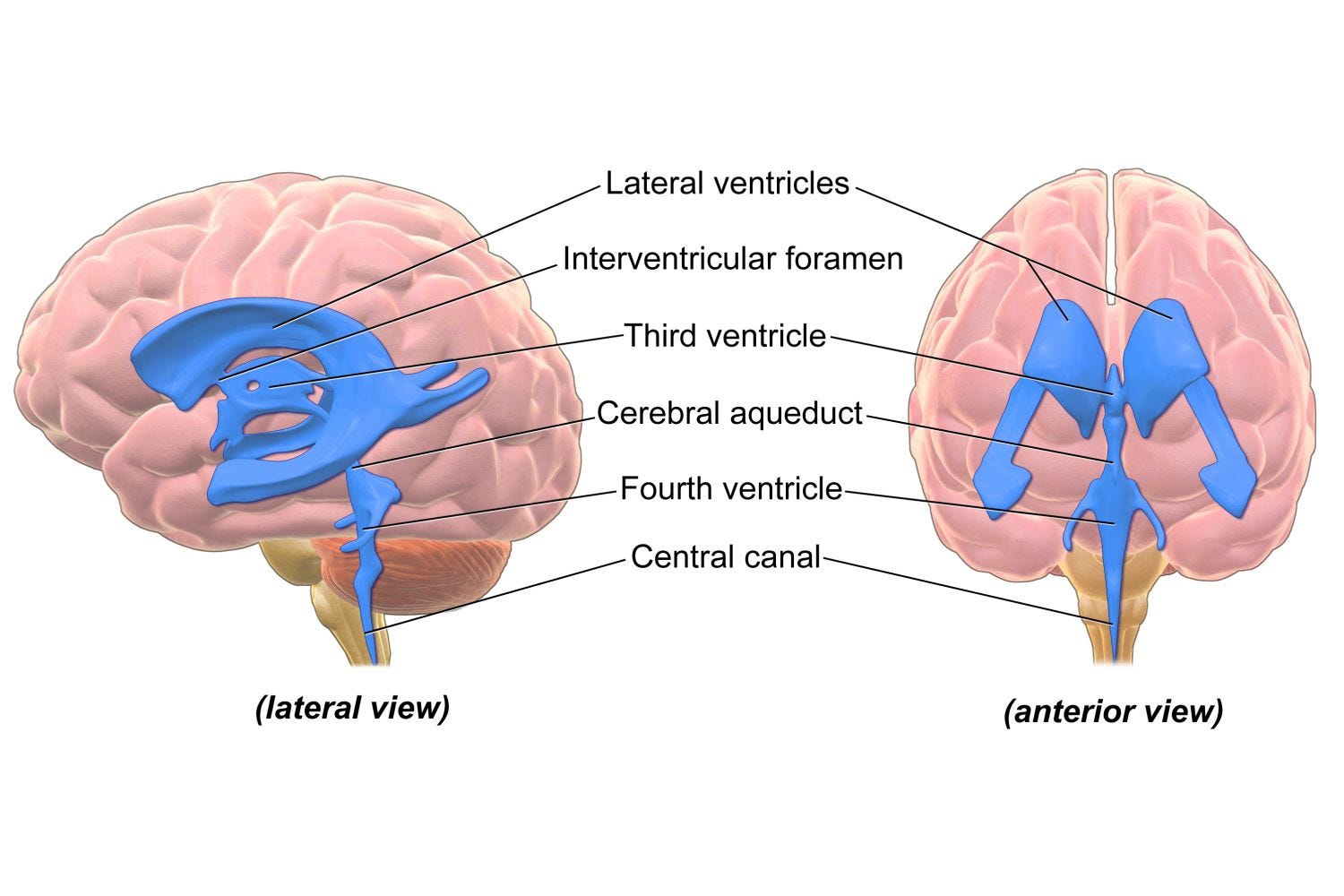
Ventricles get bigger for two structurally different reasons. In the first, fluid accumulates faster than it drains and pushes the ventricle walls outward. This is hydrocephalus, and the surrounding brain tissue gets compressed. In the second, brain tissue is lost to a degenerative or vascular process and fluid fills the space that the tissue used to occupy. Radiologists call this ex vacuo enlargement, meaning enlargement into emptiness. The ventricles look larger in both cases but the mechanism behind them is opposite.
Normal pressure hydrocephalus is the first of these two, occurring in older adults, with cerebrospinal fluid pressure that measures normal on a spinal tap. Dr. Salomon Hakim described the syndrome in the 1960s as a combination of walking difficulty, cognitive slowing, and bladder urgency, occurring together with enlarged ventricles and normal pressure. The definition has not changed much since.
How the diagnosis is currently made
A diagnosis of probable normal pressure hydrocephalus requires symptoms plus imaging. The imaging measures include the Evans index, which is the ratio of the width of the front portion of the ventricles to the width of the skull at its widest inner point, with a value above 0.3 counting as enlarged. Radiologists also look at the callosal angle, which describes how sharply the two halves of the ventricle roof meet, and at a pattern called DESH, in which the ventricles are enlarged while the grooves on the top surface of the brain are squeezed shut and the fissures on the sides are widened.
The other half of the workup is functional. Removing a volume of spinal fluid through a lumbar puncture, or draining fluid continuously for a few days, tests whether walking speed and cognition improve when the fluid load is reduced. Improvement after temporary drainage is the strongest available predictor of whether a permanent shunt will help.
How common enlarged ventricles are compared with how common the condition is
The gap between these two numbers is the central fact about this diagnosis.
Enlarged ventricles are very common in older adults. In the Gothenburg population study of 1,238 adults aged 70 and older, an Evans index above 0.3 was present in 256 people, or 20.7 percent. The squeezed grooves at the top of the brain that suggest a fluid problem rather than tissue loss were present in only 5.4 percent.
The condition itself is far less common. In the same study, probable normal pressure hydrocephalus was found in 0.2 percent of those aged 70 to 79 and 5.9 percent of those aged 80 and older. A later population-based Swedish study in Jämtland put the prevalence at 3.7 percent of adults 65 and older, with 8.9 percent in those over 80 and 2.1 percent in those aged 65 to 79. An MRI study of 791 seventy-year-olds found 1.5 percent.
Diagnosis rates are low and one reason is that most people never get a brain MRI. A study of the US National Inpatient Sample found an average prevalence of 179 per 100,000 among hospitalized patients aged 60 and older between 2007 and 2017, which is 0.18 percent. A companion analysis found an annual inpatient incidence of 2.86 per 100,000, rising to 18.81 in the 85 and older group. Community screening finds the condition in a few percent of older adults and hospital coding finds it in a fraction of a percent, which is where the underdiagnosis claim comes from.
My own view is that more older adults should be getting brain imaging and the case does not rest on hydrocephalus alone. Unruptured brain aneurysms are found in roughly 1 to 3 percent of adults. Some glioblastomas evolve from slower growing tumors that are visible on MRI for years before they produce symptoms, which is part of the broader argument I made in my article on lowering late stage cancer risk. One scan addresses all three questions.
What enlarged ventricles predict about cognitive decline
The largest dataset comes from Kuopio in Finland, where Dr. Anne Koivisto and colleagues followed 468 patients with enlarged ventricles and suspected normal pressure hydrocephalus for a median of 4.8 years. Dementia was present at follow-up in 59 percent of the whole group. Among patients who were not shunted, 73 percent developed dementia. Among those who were shunted and initially responded, 46 percent developed dementia. Dementia was 1.6X as common in the unshunted group as in the shunted responders, an absolute difference of 27 percent.
The Kuopio patients had all been referred to a hospital because someone already suspected the condition. Their conversion rate is therefore higher than an unselected sample would show. In an untreated population sample from Gothenburg followed for up to 25 years, among people with possible normal pressure hydrocephalus or asymptomatic ventricular enlargement who did not have dementia at the start, 40 percent developed dementia during follow-up, at 2.6 times the rate of people without those findings.
Enlarged ventricles without symptoms also convert. Japanese researchers have named this state AVIM, standing for asymptomatic ventriculomegaly with features of the condition on MRI. In a community sample, two of eight asymptomatic people developed walking or memory problems over four to eight years. In a hospital-based multicenter study, 27 of 52 participants, or 52 percent, developed the full condition over three years, and the DESH pattern predicted who progressed. A six-year Swedish follow-up found that 85 percent of participants with the radiological pattern alone developed symptoms. People labeled asymptomatic also test slightly below normal on word generation and motor control when measured formally. The authors interpreted this as a prodromal stage of the condition, meaning an early phase in which the underlying process is already active before symptoms become recognizable.
Why the diagnosis rests on a convention rather than a test
Alzheimer’s disease moved to a biological definition because it has a defining pathology and blood and imaging markers that track it. I have written about that shift and about what neurofilament light adds to it. Normal pressure hydrocephalus has none of these anchors.
The pressure is normal. There is no physiological threshold to measure against, which is the entire meaning of the name.
There is no defining pathology. Frontal brain biopsies taken during shunt surgery frequently show Alzheimer’s changes instead. A 2026 meta-analysis of 16 studies covering 1,825 patients found that the share with coexisting Alzheimer’s changes before shunting ranged from 23.9 to 67.6 percent across cohorts, and that this group recovered less cognitively over the long term.
The imaging is not specific. One in five older adults has ventricles above the size threshold. In the Gothenburg sample, 26 of those 256 people met criteria for probable normal pressure hydrocephalus, which is about one in ten.
The definition is partly circular. In practice, the operational test for normal pressure hydrocephalus is whether the person improves after cerebrospinal fluid is removed, and improvement is assessed by measuring symptoms. Remove symptoms from the definition and there is currently no way to identify who has it.
The symptom requirement is therefore doing the job that a biomarker does in other diseases. It separates the small number of people with a fluid disorder from the very large number with similar anatomy and no disorder. It is a threshold for intervention rather than a claim that a preclinical stage does not exist.
Because a diagnosis requires symptoms, no trial has ever placed a shunt in someone who lacks them. So there is no treated asymptomatic group whose later dementia rates could be compared against an untreated one. Everything in the literature about earlier intervention is inferred from people who were already symptomatic at the time of treatment.
Whether the atrophy causes the enlargement or the enlargement causes the atrophy
This is the question that determines whether treatment can help, and there is no clean test for it. The two processes are not mutually exclusive, since long-standing fluid pressure produces genuine and partly permanent white matter loss and degenerative atrophy coexists with a fluid problem in a large share of patients. Several features carry partial information.
The space around the outside of the brain is the most informative feature. Inside a fixed skull, tissue loss expands the ventricles and the surface grooves together. A fluid problem expands the ventricles while compressing those grooves. The disproportion between the two is what the DESH pattern captures.
The angle of the ventricle roof reflects mechanical force. Upward bowing and stretching of the corpus callosum, the band of fibers connecting the two hemispheres, indicates that the ventricle roof is being pushed outward. Tissue does not get bowed upward by its own absence.
The tissue compartment that is lost differs. Automated volume analysis comparing the two conditions found that global ventricular enlargement with marked white matter reduction and preserved gray matter characterized normal pressure hydrocephalus, while global and front-and-top-weighted gray matter reduction characterized Alzheimer’s disease.
Reversibility settles the question after the fact. Ventricles that enlarged because tissue disappeared cannot shrink. Serial imaging around shunt surgery shows that ventricles do become significantly smaller afterward, in patients who improve clinically and in those who do not.
The white matter changes that accompany enlarged ventricles are part of the same question. Brain scans in older adults frequently show bright patches in the white matter, which is the wiring layer beneath the surface of the brain that carries signals between regions. These patches are called white matter hyperintensities and they are the most common imaging marker of small vessel disease, meaning damage to the smallest blood vessels in the brain. A larger burden of them is associated with higher risk of stroke and cognitive decline in the general population.
The patches immediately surrounding the ventricles do reflect the fluid problem, and three separate lines of evidence support that. Injecting contrast into the spinal fluid shows that it migrates across the ventricle lining into the surrounding white matter and that the degree of signal change tracks how much contrast arrives there. Diffusion imaging finds that free water content in those regions is elevated compared with controls. And their width is reduced after shunt surgery in the patients who improve, which is what reabsorbing fluid would do and what established tissue damage would not. They do not establish elevated pressure, since the pressure measures normal by definition, and the direction of the flow is contested because the same tracer work shows fluid moving inward from the surface as well. The leading mechanistic account describes fluid crossing the ventricle lining as an alternative drainage route recruited because normal drainage is impaired rather than as a consequence of high pressure. The lesions further from the ventricles behave differently, since one study using a measure of fluid clearance found that the marker correlated with ventricular volume but not with total white matter lesion volume, and those deeper lesions are considered primarily ischemic and occur in both conditions.
What treatment involves and what it delivers
The treatment is a shunt, a thin tube placed from a ventricle under the skin to the abdomen, with a valve that controls how much fluid drains.
The gait evidence is now randomized. The PENS trial, published in the New England Journal of Medicine in September 2025 and led by Dr. Mark Luciano, Dr. Michael Williams, and Dr. Mark Hamilton, enrolled 99 patients across 17 centers in the United States, Canada, and Sweden. Everyone received a shunt. Half had the valve set open and half had it set to a pressure high enough that essentially nothing drained, and neither patients nor assessors knew which. At three months, walking speed and a combined gait and balance measure improved significantly in the open group. This resolved a long-running question about whether earlier results reflected the placebo effect of surgery.
The cognitive result was negative at three months. Scores on the Montreal Cognitive Assessment improved by 1.3 points with an open shunt and 0.3 points with a placebo setting, which was not a significant difference. Bladder symptoms also showed no difference. The investigators note that cognitive changes have long been suspected to take longer and require more detailed testing, and the trial is following participants to twelve months.
The risks are real. In PENS, subdural bleeding occurred in 12 percent of the open shunt group compared with 2 percent of the placebo group, and positional headaches in 59 percent compared with 28 percent. Falls went the other way, occurring in 24 percent of the open shunt group and 46 percent of the placebo group. In a Norwegian series of 227 adults followed for one to eleven years, 20.7 percent required revision surgery, 45.4 percent had at least one complication, and 8.2 percent of procedures involved a severe or fatal complication.
The dementia prevention evidence is observational. An analysis of 2,053 patients that statistically matched treated and untreated groups from the Taiwan National Health Insurance Research Database found that shunted patients had a lower subsequent risk of dementia, at about three quarters the rate of the unshunted group, and a lower risk of Alzheimer’s disease. The range of values statistically compatible with the dementia result stopped just short of no effect at all. Vascular dementia showed no difference. The Alzheimer’s estimate rests on few events, only 376 patients were shunted, and treatment assignment in a database selects for younger and healthier patients even after statistical adjustment.
Putting the numbers together, if the condition accounts for one to five percent of dementia and the observed relative reduction holds, the arithmetic lands somewhere between roughly 20,000 and 100,000 preventable cases against a US dementia population above seven million.
How the calculation changes if people live to 120 rather than 80
Much of the conservatism around shunting older adults comes from competing mortality. When five-year all-cause mortality in untreated probable normal pressure hydrocephalus is near 88 percent, a large share of patients do not survive long enough for a dementia diagnosis either way and a surgical complication rate near 8 percent yields very little over a short remaining lifespan.
If a healthy 80-year-old has 25 or 40 years ahead rather than 6, several inputs move. The years of preserved mobility gained from a successful shunt multiply. The cumulative cost of untreated gait failure, falls, and loss of independence grows. A one-time surgical risk amortized over four decades looks different from the same risk amortized over five years. On that logic, a fit and independent 80-year-old with clear imaging features and a good response to temporary drainage is a stronger candidate than the frail 80-year-old with multiple conditions, which is the reverse of how age alone is often used.
The same structure appears in cancer screening. Concern about overdiagnosis, meaning treatment of tumors that would never have caused harm, carries different weight depending on how long someone lives and I have made that argument elsewhere.
The evidence for a dementia benefit is observational rather than randomized. Both the Kuopio comparison and the Taiwanese cohort point toward fewer dementia diagnoses after shunting and preventing dementia is a cognitive benefit by any definition, but neither study randomized who received a shunt. A longer lifespan multiplies whatever the true effect turns out to be. It also multiplies the accumulated risk of shunt complications, since revision rates in the Norwegian series continued over eleven years of follow-up, and a shunt placed at 80 in someone expected to reach 120 is a device that has to keep working for four decades.
The gait benefit is the one supported by randomized trial evidence and it compounds over every additional year of life. Falls were also less frequent in the open shunt group, though that came from adverse event reporting rather than a prespecified endpoint.
What would need to be built to answer these questions
A biological marker that does not depend on symptoms. Candidates include measures of fluid clearance based on how water moves through tissue, where one study separated patients from controls almost perfectly, and imaging that distinguishes loose fluid from established damage in the white matter. Neither has been validated prospectively at scale.
A predictor of who converts. The DESH pattern is the best current candidate and it predicted progression in the multicenter study. The entire evidence base for conversion rests on cohorts of 8 to 52 people with estimates ranging from 25 percent over eight years to 17 percent per year.
Better selection of who gets treated. The arithmetic for a prevention trial is closer than it first appears. The observational data imply an absolute dementia risk reduction somewhere around 10 to 15 percent, against an 8 percent rate of severe or fatal surgical complications. Genuine uncertainty about which side wins is the condition under which a trial is justified. What determines whether the ratio works is who gets enrolled, since enlargement caused by tissue loss does not respond to drainage. In the Gothenburg sample, 56 of the 256 people with enlarged ventricles had imaging consistent with hydrocephalus, and the DESH pattern narrows the group further to those who go on to convert. The Johns Hopkins group has stated it is working on less invasive diagnostic methods and entry criteria of that kind are what would make such a trial feasible.
A trial with a reachable endpoint. Conversion from asymptomatic imaging findings to symptomatic disease accumulates within three to six years and would power a study in hundreds of participants. A dementia endpoint is also within reach in this population, since baseline dementia rates between 40 and 73 percent put the requirement in the hundreds to low thousands depending on the effect size assumed, far below what a general population prevention trial would need. The harder constraint on a dementia endpoint is follow-up duration and the competing risk that participants do not survive long enough to reach the outcome.
What this means in practice right now
For someone whose brain MRI shows enlarged ventricles and who has no symptoms, serial imaging to establish whether the ventricles are growing tells more than any single measurement.
Formal testing matters more than a symptom history here, because people classified as asymptomatic on questioning frequently show measurable deficits when walking speed and skills like planning and mental flexibility are quantified. That testing establishes a baseline against which future change can be measured, which is what converts an ambiguous scan into a decision.
For someone who does have walking difficulty, cognitive slowing, or bladder urgency alongside enlarged ventricles, the evidence supports evaluation by a neurologist or a center with a dedicated hydrocephalus program, including a trial of temporary fluid drainage. Response to that trial remains the best available predictor of who benefits, and it was the enrollment criterion for the one randomized trial that has shown a benefit.
The periventricular white matter changes that accompany enlarged ventricles do respond to drainage. In a series imaged before and after surgery, their width was reduced in the patients who improved and the degree of reduction correlated with clinical improvement. Blood pressure control and the other established modifiable risk factors I have covered elsewhere address the vascular contribution separately.
The question that is unsettled for me is what someone should do who has mild symptoms, imaging features that fit, a good response to temporary drainage, no other serious illness, and an expectation of living another twenty or thirty years. For that person the gait benefit is established, since those are close to the criteria PENS enrolled on. The dementia benefit is supported by observational data and has not been tested in a randomized trial and the trial that would settle it does not currently exist. Waiting for it means accumulating years of untreated disease in the person who stands to gain the most from a long remaining lifespan.
If it were me, I would take the shunt because I am more worried about dementia than about mortality. Someone reading the same numbers could reasonably decide otherwise, given the 8 percent rate of severe or fatal complications with shunt surgery.
More people should be screened for normal pressure hydrocephalus and followed over time. The symptoms overlap with common features of aging and the diagnosis depends on imaging that most people never receive. A brain MRI obtained for this question also finds unruptured aneurysms and slow growing tumors. It establishes a baseline against which white matter changes can be tracked over time. Most of the uncertainty in this article concerns asymptomatic and ambiguous cases. The people who have symptoms, imaging that fits, and a clear response to temporary drainage face a much more straightforward decision, and they are the ones most often going undiagnosed.


Written by
Dr. Christin Glorioso, MD PhD
Dr. Glorioso is the founder and CEO of NeuroAge Therapeutics. With her background in neuroscience and medicine, she is dedicated to revolutionizing brain health and helping people maintain cognitive vitality.
Learn more about Dr. Glorioso

