
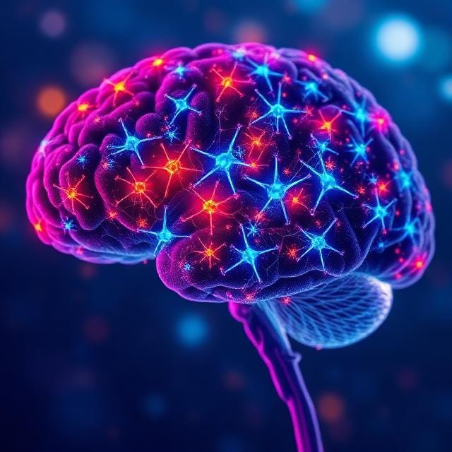
Does the adult human brain make new neurons? (new study out in Nature)
People are able to increase the volume of their brain on MRI in clinical studies through exercise and other interventions, which is remarkable. An open question is whether that increase volume is because new neurons are being born, or if it instead reflects the neurons becoming “fluffier” (having more synaptic connections between neurons), or both.
A paper published in Nature in February 2026 by Dr. Orly Lazarov and colleagues at the University of Illinois Chicago, Northwestern University, and the University of Washington offers the most detailed molecular look into this questions, which is one of the most debated questions in brain science. Does the adult human brain keep producing new neurons through a process called neurogenesis (the production of new neurons from stem cells), and if so, what happens to that process as we age and as Alzheimer’s disease takes hold? Using gene-reading technology (RNA seq) applied to nearly 356,000 individual brain cells from 38 donated human brains, the study provides additional evidence that neurogenesis occurs in the adult human hippocampus, shows how it breaks down in Alzheimer’s disease, and identifies a distinctive molecular pattern in people who maintain exceptional memory well into old age.
Whether the adult human brain generates new neurons has been debated for decades. For much of the twentieth century, the dominant view was that the brain you are born with is largely the brain you keep. That changed in 1998, when a study by Dr. Peter Eriksson, Dr. Fred Gage, and colleagues used a DNA-labeling technique in cancer patients to show that new neurons were being born in the hippocampus of adult humans. The hippocampus is the brain region most involved in forming new memories, and the idea that it could keep producing neurons into old age had obvious implications for how we think about learning, cognitive aging, and dementia.
The story continued in 2018, when two papers were published with opposing conclusions. A study in Nature led by Dr. Shawn Sorrells found no detectable young neurons in the adult human hippocampus, concluding that neurogenesis drops to undetectable levels by adulthood. Within weeks, a study in Cell Stem Cell led by Dr. Maura Boldrini came to the opposite conclusion, reporting thousands of immature neurons in tissue from healthy adults up to age 79. The field has been working to reconcile those findings ever since. The 2026 Lazarov paper is the latest contribution to that effort.
Why earlier studies disagreed with each other
The disagreement was largely about methodology. In living rodents, scientists can inject chemical tags that mark cells as they divide, then count the tagged cells weeks later. That approach cannot be used in healthy living humans for research purposes. It was only possible in the 1998 study because those participants were already receiving BrdU (the chemical tag for dividing cells) as part of cancer treatment. Instead, researchers used protein markers that appear on young neurons in postmortem brains, but those markers behave differently in human tissue than in rodent tissue, and they are sensitive to how brain tissue is handled after death. A 2019 study by Dr. Maria Llorens-Martin’s group in Madrid showed that leaving tissue in preservative solution for too long nearly eliminated the relevant protein signal entirely. The Sorrells samples spent longer in preservative, which likely accounts for at least part of the discrepancy with studies that did find neurogenesis.
Rather than relying on a single marker protein, the 2026 study read the gene activity of nearly 356,000 individual brain cells and used a second technique to measure how each cell’s DNA was physically organized and regulated. Both measurements were taken in the same cells simultaneously, and the results could be checked against independent datasets from other labs. This makes the approach considerably more resistant to the technical problems that affected earlier work.
What the 2026 study showed
The researchers examined brain tissue donated after death from five groups of people. The first was eight cognitively healthy adults between ages 20 and 40. The second was eight older adults with no cognitive impairment, representing typical healthy aging. The third was six adults who had early Alzheimer’s-related changes in their brain tissue but had not yet been diagnosed. The fourth was ten adults with diagnosed Alzheimer’s disease. The fifth, and most novel, was six SuperAgers, defined as adults aged 80 or older whose memory tested as well as people in their 50s.
For each cell, the team measured two things simultaneously. The first was which genes were actively switched on (single-nucleus RNA seq), giving a snapshot of what the cell was doing. The second was which parts of the cell’s DNA were physically open and readable (snATAC-seq), reflecting deeper regulatory controls. Together, these two readouts allowed the researchers to identify what kind of cell each one was, and to trace the molecular path it was moving along.
A developmental pathway from stem cells to mature neurons is present in healthy adult brains
In the young adult brains, the researchers found stem cells, precursor cells, and immature neurons, and were able to show that cells were progressing in a clear direction along that sequence, from stem cell toward mature neuron. Each stage had a distinct molecular signature that matched what would be expected if genuine neurogenesis were occurring. Two independent datasets from other labs showed the same pattern, and when the team tested their signature against brain regions where new neuron production would not be expected, it did not appear there, suggesting the signal reflects real biology rather than a measurement artifact.
In Alzheimer’s disease, the process appears to get stuck rather than simply shut off
In the Alzheimer’s group, precursor cells and immature neurons were significantly less abundant than in healthy brains. Neural stem cells, however, were not depleted; they were more abundant. The most straightforward interpretation is that stem cells are still present but cannot complete the journey to becoming mature neurons. The process stalls midway. This pattern was consistent with several earlier studies using different methods, including Tobin et al. (2019), Moreno-Jimenez et al. (2019), and the Zhou et al. 2022 Nature paper, which also found reduced immature neurons in Alzheimer’s tissue.
The reason why stem cells would stall in Alzheimer’s are not fully established, but candidate mechanisms include the toxic effects of amyloid and tau on differentiating cells, chronic neuroinflammation, reduced BDNF, and the disrupted DNA regulatory programs the paper itself identified in early-stage disease.
Changes in how DNA is packaged appear before changes in gene activity in people with early Alzheimer’s pathology
One finding with potential implications for early detection concerns the preclinical group. These individuals had early Alzheimer’s-related changes in their brain tissue but had not been diagnosed during their lifetimes. When the researchers looked at which genes were active in their neurogenic cells, the pattern was relatively similar to healthy older adults. When they looked at how the DNA was physically organized and regulated, however, substantial differences were already present compared to all other groups.
This matters because it suggests molecular disruption in neurogenic cells may begin earlier than previously recognized. Changes in DNA organization appear to precede changes in gene activity. Whether this could eventually be detected from blood or spinal fluid as an early warning sign is an open question, but the finding points in that direction.
SuperAgers show a molecular profile in their young neurons that resembles younger brains
SuperAgers (people with cognition like someone 30 years younger) had more precursor cells and immature neurons than the Alzheimer’s group.
This is consistent with my 2019 paper showing people with younger looking brains by gene reading (RNA seq) are much less likely to have Alzheimer’s and have better memories.
The researchers also identified a molecular signature that was stable across young adults, healthy older adults, and SuperAgers but reduced in Alzheimer’s disease, particularly in the regulatory packaging around DNA. Among the genes elevated in the young neurons of SuperAgers was BDNF (brain-derived neurotrophic factor), a protein that supports neuron survival and the strengthening of connections between neurons, which I have written about in the context of exercise and brain health.
The gene regulatory programs active in SuperAger stem cells and immature neurons overlapped more with those of young adults than with typical healthy older adults. This suggests that SuperAging may involve maintaining a more youthful molecular environment in neurogenic cells, rather than simply aging more slowly. The SuperAger group was small, only six individuals, so these findings should be treated as preliminary until confirmed in larger studies.
Broader hippocampal differences also separate those who age well from those who do not
Beyond the neurogenic cells, other cell types in the hippocampus also differed between groups. The neurons responsible for memory consolidation and retrieval showed the largest differences in gene activity between SuperAgers and people with early Alzheimer’s pathology, with many of the most changed genes involved in how neurons communicate with each other. The support cells of the brain showed pronounced differences in DNA organization between groups, suggesting that the molecular environment of a resilient hippocampus involves coordinated changes across multiple cell types, not just those producing new neurons.
Several important limitations apply to these findings
The sample sizes are small. The SuperAger group had only six individuals, and the authors note that this limited their ability to detect differences in cell abundance with confidence. The cells involved in neurogenesis are rare within the hippocampus as a whole, which makes precise counting difficult.
The study also cannot confirm whether the immature neurons it identifies go on to mature and become functional. In rodents, newly born neurons integrate into circuits and contribute to memory. Whether the same occurs in adult humans, and at what rate, is still unknown. The presence of immature neurons is consistent with ongoing neurogenesis but does not prove those cells complete the process, in healthy aging or in disease.
The molecular patterns identified here are associative rather than causal. Showing that a particular regulatory program is present in SuperAger stem cells does not prove that program is responsible for their cognitive resilience. Testing causality would require different kinds of experiments.
This study moves the neurogenesis debate forward without fully resolving it
The broader debate about adult human neurogenesis has shifted since 2018, though it has not resolved. The existence question remains contested, and serious researchers still dispute whether meaningful neurogenesis occurs in the adult human hippocampus at all, but the weight of evidence has continued to move toward the view that it does. The downstream questions about how much occurs, what regulates it, and what role it plays in memory and disease are largely still open. The 2026 Lazarov paper adds meaningful evidence on all of these fronts, particularly by showing that molecular changes in the packaging around DNA track cognitive status more closely than changes in gene activity alone.
It also addresses the methodological concerns raised by the Sorrells paper. By comparing its findings against both the Frisén 2025 Science dataset and brain regions where neurogenesis would not be expected, the study makes a technically stronger case for the presence of neurogenic cells than prior work.
What this means for lifestyle and brain health
This study cannot tell us how to protect our own neurogenesis. It shows that neurogenesis looks different across cognitive groups but cannot establish what caused those differences. That said, the molecular features that distinguished the resilient hippocampus in this dataset overlap with what other research has shown about the effects of lifestyle on brain health.
Exercise has the clearest evidence in animal models. Aerobic activity raises BDNF levels and directly increases new neuron production in rodent hippocampi, and BDNF was elevated in SuperAger immature neurons in this study. I have written about exercise and brain health in more detail here. The communication pathways between neurons that were better preserved in SuperAgers and healthy older adults are also supported by cognitive engagement and social activity in observational studies of human aging. Sleep quality matters too, because the brain’s waste clearance system, which operates primarily during sleep, has downstream effects on hippocampal neurogenesis and synaptic health. I have covered the glymphatic system and sleep in more detail here.
The Mediterranean diet has the strongest human observational evidence linking diet to hippocampal volume and cognitive aging, with several studies associating higher adherence with slower hippocampal atrophy and reduced dementia risk. The energy metabolism and synaptic pathways identified as features of a resilient hippocampus in this paper overlap with pathways influenced by diet, which makes the connection plausible, but plausibility is not evidence. I have written about diet and brain health in more detail here.
Why the mechanism behind exercise-induced hippocampal growth is not yet settled
When aerobic exercise increases hippocampal volume in humans, as shown in randomized controlled trials including a 2011 study by Erickson and colleagues in which 120 older adults showed approximately 2% growth in the front portion of the hippocampus after one year of aerobic walking, the question of what is actually growing inside the hippocampus is not fully understood. The volume change measured by MRI reflects several things happening at once, including the birth of new neurons, the growth of branches on existing neurons, increases in the number of connections between neurons, the formation of new blood vessels, and changes in supporting brain cells. Standard MRI cannot tell these apart.
In rodents, neurogenesis contributes meaningfully to exercise-induced hippocampal volume gains. Studies that selectively block new neuron production using radiation or genetic methods reduce a portion of the volume effect but not all of it, which shows that growth of existing neurons, formation of new blood vessels, and other processes are also happening. Even in rodents, where exercise-induced neurogenesis is well established, the volume increase has more than one cause.
In humans, the Erickson trial found that blood levels of BDNF were linked to the hippocampal volume increase, which is consistent with a neurogenic interpretation given BDNF’s role in supporting new neuron survival and maturation. But BDNF also strengthens existing connections between neurons and promotes the growth of branches on existing cells, independent of neurogenesis, so BDNF levels alone cannot confirm which process is driving the volume change. The 2026 Lazarov paper is relevant here. It confirms that the biological machinery needed to produce new neurons, including stem cells, young precursor cells, and immature neurons, is present in adult human hippocampi, and that BDNF is elevated in the most cognitively resilient group. This means the capacity to respond to exercise is present. Whether it does respond to exercise in humans, and whether that accounts for the volume changes seen in controlled trials, remains an open question.
The imaging and biomarker tools that could eventually answer this question
Directly observing neurogenesis in a living human brain requires methods that can identify individual cells or small populations of cells deep within the hippocampus without surgery or biopsy. No currently available imaging tool can do this, but several approaches are in development or have been tested in animals.
The most developed non-invasive approach uses PET scanning (positron emission tomography, a type of brain scan that detects radioactive tracers taken up by specific cells) with a compound called [18F]FLT. This compound gets incorporated into cells that are actively dividing, so in principle it can tag stem cells in the process of producing new neurons. The practical problem is that the brain’s protective barrier between blood and brain tissue actively pumps this tracer back out before it can accumulate in high enough concentrations to produce a clear signal. Researchers at RIKEN in Japan addressed this by giving rats probenecid, a gout medication that blocks the proteins responsible for pumping the tracer out, substantially increasing accumulation in neurogenic brain regions. Using this approach, the team produced PET images that could distinguish between reduced neurogenesis under stress and recovery with antidepressant treatment, confirmed by examining brain tissue directly afterward. Both probenecid and the tracer are in principle usable in humans, and the RIKEN group reported beginning tests in non-human primates, but human validation has not been published.
Even if this PET approach were validated in humans, it would detect cell division rather than confirm that the dividing cells are neurons. Many types of cells in the hippocampus divide, including support cells and cells lining blood vessels, not only the precursors to new neurons. To get around this, researchers would need a tracer that binds specifically to proteins found only on young neurons during their development. One candidate target is the protein called doublecortin, discussed earlier in this article as a marker for immature neurons. No such tracer has yet been validated for use in humans.
Blood and spinal fluid biomarkers offer a complementary but indirect approach. BDNF in blood rises reliably with aerobic exercise in humans and indicates that the hippocampus is responding to activity in some way, but it reflects a broad range of brain changes rather than neurogenesis specifically. Other proteins more closely tied to the production of new neurons, including doublecortin and a related molecule called PSA-NCAM that appears on young neurons as they mature, have been measured in the spinal fluid of animal models as potential markers for neurogenic activity. Whether these proteins are reliable enough in living humans has not been established.
The most definitive study design would use a validated neurogenesis-specific PET tracer in a randomized controlled exercise trial, measuring how much new neuron production is occurring before and after the intervention. That design is feasible in principle. The missing piece is a human-validated PET tracer that can reliably pick out neural stem cells and their progeny from the surrounding tissue. Until one exists, the cellular basis of exercise-induced hippocampal growth in humans will remain an inference from animal studies rather than a directly observed phenomenon.
This is a question that comes up in the context of brain MRI assessments, including the volumetric MRI that NeuroAge uses to track hippocampal size over time. When someone’s hippocampal volume increases between scans, the MRI cannot tell us what drove that change. New neurons, new dendritic branches on existing neurons, new synaptic connections, new blood vessels, and changes in supporting cells can all contribute to a larger measured volume.
The finding from this paper that the biological machinery for neurogenesis is present in adult human hippocampi, and that BDNF is elevated in the most cognitively resilient group, is consistent with neurogenesis playing a role. For now, the most accurate answer to the question of what is growing is that we know something structural is changing, which is likely new neurons at least in part, and we have good reason to think lifestyle factors drive part of that brain growth.
Why does knowing what drives brain growth matter? We’d like to be able to mimic the factors that cause brain growth with a therapeutic or intervention to give everyone new neurons to stay sharp and treat Alzheimer’s. In order to do that, it’s helpful to understand the process that produces brain growth naturally. This new study adds to the information that we need to make these future therapies a reality.

Written by
Dr. Christin Glorioso, MD PhD
Dr. Glorioso is the founder and CEO of NeuroAge Therapeutics. With her background in neuroscience and medicine, she is dedicated to revolutionizing brain health and helping people maintain cognitive vitality.
Learn more about Dr. Glorioso

